Platform

Biochemistry and Structural Biology

Key Techniques

– Expertise in protein expression and purification, enzyme kinetics,
assay development, high-throughput screening (HTS), target
engagement, BioID, protein interactions.
– HTS to identify hit compounds that are tested in orthogonal
biochemical and phenotypic screening assays.
– Target engagement with Differential Scanning Fluorimetry (DSF),
Drug Affinity Responsive Target Stability (DARTS) and Cellular
Thermal Shift Assay (CETSA).
– Collaboration with Professor Pål Stenmark (since 2011) for
crystallization of co-complexes to guide structure-based design of
potent inhibitors and determination of protein 3D structures.

The biochemistry, assay development and in vitro pharmacology team holds vast expertise in the areas of protein expression and purification, enzyme kinetics, assay development, high-throughput screening and target engagement analysis methods.

After target validation we produce expression constructs for eukaryotic and prokaryotic production of the protein of interest. Target proteins as well as proteins needed for determining the selectivity of inhibitors are generally produced using recombinant protein expression in E. coli, followed by purification using state-of-the-art methods comprising affinity purification, ion exchange chromatography and size exclusion chromatography. The purity of protein preparations are carefully assessed using SDS-PAGE gel analysis.

We develop assays suitable for high-throughput screening (HTS) to monitor enzyme activity, protein binding and inhibitor potency of compounds. Assay performance is thoroughly evaluated and Z’-factor as well as assay window is carefully monitored.

We perform HTS of small molecule libraries in the search for enzyme inhibitors or protein binders and perform hit confirmation and counter screening to filter out false positive and unselective compounds. Hit compounds are tested in orthogonal biochemical and phenotypic screening assays. We perform HTS screening of fragments using Differential Scanning Fluorimetry (DSF). We collaborate closely with the SciLifeLab Compound Center for plating of compound libraries.

To assess target engagement in vitro we routinely run DSF and nano-DSF assays. In cells we use Drug Affinity Responsive Target Stability (DARTS) and Cellular Thermal shift assay (CETSA). The team works closely with the medicinal chemistry platform in early drug discovery projects that we run in the lab through reiterative directed compound synthesis and activity screening to improve compound properties.

We closely collaborate with Professor Pål Stenmark at Stockholm University and his co-workers on crystallization and determination of 3D protein structures and of co-complexes with compounds to guide structure-based design of potent inhibitors.

To elucidate the cellular function of proteins we perform enzyme kinetic experiments and BioID methodology to identify protein interactions as they occur in living cells. We also perform protein interaction analysis using co-immunoprecipitation followed by western blotting or mass spectrometry analysis. We use site-directed mutagenesis to generate tools to further understand protein function and to identify amino acid residues crucial for enzyme activity as well as inhibitor potency.

Medicinal Chemistry and Pharmacology

– Target selection and prioritization, of assay development, selection of screening libraries, screen-to-hit activities such as evaluation and triaging of high-throughput screening results, hit expansion and lead optimization strategies, design, and synthesis of fluorescent probe compounds, scale-up of compounds for pharmacokinetic and pharmacodynamic studies in animal models and patent strategies.
– Screening funnel and determination of ADME (absorption-distribution-metabolism-excretion) properties in collaboration with UDOPP and in vitro and in vivo pharmacology.

The medicinal chemistry team in the Helleday Laboratory is comprised of a mix of scientists with backgrounds from both academic and industry drug discovery.

Medicinal chemistry is integral to most aspects of research within the lab. Our team are routinely involved in target selection and prioritization, assay development, selection of screening libraries, screen-to-hit activities such as evaluation and triaging of high-throughput screening results, hit expansion and lead optimization strategies, design and synthesis of fluorescent probe compounds, scale-up of compounds for pharmacokinetic and pharmacodynamic studies in animal models and patent/publication strategies.

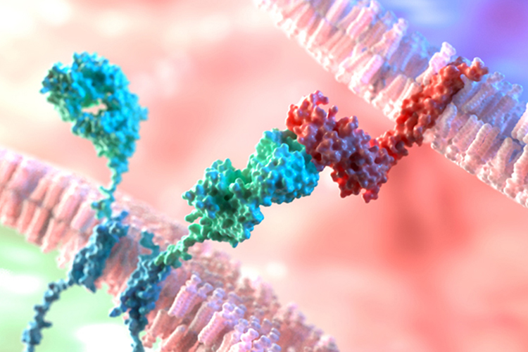
We have developed many first-in-class potent inhibitors against novel protein targets within the lab. Some examples are: MTHFD1/2, MTH1, OGG1, NUDT5, dCTPP1, NUDT15, NUDT22, and SAMHD1. In our OGG1 program we have also developed extremely interesting ‘amplizyme’ compounds (activators) capable of causing a gain-of-function in the OGG1 protein and increasing its biochemical activity. Notably the dual action MTH1/tubulin inhibitor program is entering phase 2 studies while both OGG1 and MTHFD1/2 inhibitor projects are in late preclinical phase preparing for first-time-in-man studies .

Screening strategies – we run a mixture of virtual screening, fragment-based approaches and traditional high-throughput screening campaigns. These are run using in-house collections or drawing from our large collaborative network including Chemical Biology Consortium Sweden and the European Lead Factory for high-throughput screening or the Carlsson Laboratory for virtual fragment screening. We have succeeded with ligand-based approaches (e.g., MTHFD2 project).

Screen-to-hit activities – often the results of high-throughput screening campaigns can be misleading and give rise to false positive hits. These can be attributed to many factors such as aggregation, unusual redox behaviour, metal chelation or intrinsically fluorescent or reactive compounds. Our chemistry team rigorously triage hits to eliminate undesirable compounds and ensure the hits we work on are verified for purity and identity. Hit compounds are usually re-synthesized and investigated in various counterscreen and biophysical assays to ensure they engage the target protein and do not interfere in the primary biochemical assay. Analogue mining in internal and external collections often allows us to identify interesting compounds to support this activity. Typically we will investigate solubility of promising hits through our ADME (absorption-distribution-metabolism-excretion) collaboration with UDOPP and collaborate with the structural group of Pål Stenmark (Stockholm University) for x-ray cocrystal information.

Hit expansion activities – medicinal chemistry in the Helleday lab has access to powerful synthetic capabilities which allow rapid analogue synthesis and investigation of SAR. We design and prepare compound arrays and matched pairs using structural information generated by crystallographic and computational approaches. Virtual libraries can be enumerated and docked and physicochemical properties such as cLogP or solubility calculated to inform decision making.

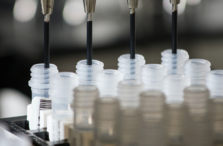
Medicinal chemistry is well served by facilities for parallel synthesis and purification such as preparative MPLC, HPLC, SFC and parallel evaporation, including walk-up LCMS and NMR analysis. In our chemical store, more than 18,000 bar-coded reagents and building blocks can be found and searched by substructure searches or CAS number.

Lead optimization – once a hit series is confirmed and SAR is established there are usually many parameters that require optimization before it could be considered as a lead or probe compound. In this phase we attempt to improve compound potency, target selectivity and pharmacokinetic properties to identify lead compounds (and their negative controls). Optimized leads can be used by our in vivo pharmacology team in relevant animal disease models to provide proof-of-principle and establish whether the protein target of interest could be of potential therapeutic interest. Many molecular parameters need to be carefully balanced to ensure that sufficient exposure is reached at the site of action. In addition to on-target potency and selectivity towards off-targets, ADME parameters such as solubility, metabolic stability, plasma protein binding, passive cell permeability and efflux may also need to be optimized simultaneously. This is done by iterative DMTA (design-make-test-analyze) cycles.

During this phase we often need to develop efficient synthesis of compounds in multi-gram scale for animal studies or develop sophisticated tagged molecules to allow us to visualize them in cells so that mechanistic insights can be gained.

In the lead optimization phase a decision is reached on whether the chemistry will be published or whether the group will first seek patent protection before publication and the project progressed towards clinical studies in man.

Disease Models

In vivo pre-clinical drug development studies in the field of inflammation, autoimmune diseases, immuno-oncology, and cancer. We perform pharmacodynamics (PD) studies in mice. We optimize maximum tolerated dose (MTD) and therapeutic dose of new small-molecule inhibitors by considering pharmacokinetics (PK) profiling, drug tolerance study, hematological and toxic-pathological analysis. We validate new cancer targets in rodent models, determine target engagement and specificity of inhibitors in tumors using CETSA. We utilize primary human tissue from patients to study the DDR. From clinical studies, we isolate biopsies and utilizes classical histological methods for tissue handling, staining and multiplexed immuno-based approaches in automated epifluorescence and confocal microscopy settings to determine the quantity of proteins, their localization, and phosphorylation status in tissues. We use Spatial Transcriptomics to achieve a full expression profile with a near single cell resolution on tissues without distorting the tissue architecture.

Fig 1 Spatial microarrays. ST procedure: matrices with read counts for every gene in every spot which are then, by an unsupervised machine learning algorithm, resulting in a set of factors (cell types, cancer, inflammation, etc.), each comprising one activity map and one expression profile.

Today we know that tumors often show vast heterogeneity in genetic alterations, gene expression patterns, and protein contents. This heterogeneity also exists within individual tumors. To gain a deeper understanding of the biology of tumors and find out strategies to eliminate them, studies on cancer cell lines will not give us the whole picture, we also need to study tumors in their natural environment, i.e., in tissue samples with tumors, taken from humans or animals.

The platform utilizes classical histological methods for tissue handling, staining and multiplexed immuno-based approaches in automated epifluorescence and confocal microscopy settings to determine the quantity of proteins, their localization, and phosphorylation status in tissues. In collaboration with Prof. Joakim Lundeberg’s group at Scilifelab we use Spatial Transcriptomics (ST) to achieve a full expression profile with a near single cell resolution on tissues without distorting the tissue architecture, making it possible to get morphological properties and annotations of pathologists from the same tissue section (Fig 1). Using laser capture microscopy or manually dissecting small tissue areas from consecutive tissue sections, we can get spatial genetic information from the same tumor.

Thus, by creating a spatial landscape of genetics, gene expression, protein properties and morphological data from consecutive tissue sections within a single tumor, we can rigorously study the interplay between the tumor and surrounding stroma between different subareas in the tumor. This approach creates large data volumes, and we have extensive collaboration with bioinformaticians to develop new tools for handling and analyzing the data. Both classical and unsupervised machine learning approaches are used (Fig 2).

Fig 2 Color coded expression signatures achieved from an unsupervised machine learning approach. A) Prostate tissue. B) Needle biopsy.

The platform analyzes tissues derived from human cancer patients that have been enrolled in different studies with the aim to better understand the complex interplay between tumor and stromal cells, as well as the significance of different subpopulations within the same tumor for development of drug resistance. We also analyze material from mouse xenografts exposed to different candidate compounds developed by the Helleday lab to study their efficacy in of human cancers. Further, we analyze biopsies from patients enrolled in clinical trials testing candidate drugs developed in our lab, to monitor efficacy and to find relevant biomarkers.

Molecular Biology of DDR

Key Techniques

– Detailed investigation of the basic biological functions of targets and interactions in live cells.
– Expertise in advanced live cell confocal microscopy in combination with laser microirradiation, Fluorescence Recovery After Photobleaching (FRAP) and Fluorescence Loss In Photobleaching (FLIP) to visualize high spatiotemporal details and highly coordinated processes of DNA repair, cell division and genome duplication in live cells.
– Bespoke modified comet assays, DNA fibre assays, Quantitative Image Based Cytometry (QIBC), siRNA and shRNA mediated knockdown, CRISPRCas9.

A deep understanding of the molecular mechanisms of DNA repair and nucleotide metabolism is the cornerstone of our drug discovery approach. To be able to efficiently and successfully target proteins involved in these pathways for the treatment of cancer and other diseases, we need a detailed view of their biological functions and interactions in live cells. We have the unique ability to use small molecule inhibitors and activators developed by our excellent medicinal chemistry team as probes in our advanced molecular and cell biology assays to uncover new important biology.

We regularly employ advanced live cell confocal microscopy in combination with laser microirradiation, Fluorescence Recovery After Photobleaching (FRAP) and Fluorescence loss in Photobleaching (FLIP) to visualize high spatio-temporal details and highly coordinated processes of DNA repair, cell division and genome duplication in live cells. This is complemented by traditional antibody-based immunofluorescence assays in tumors and cells, allowing visualization of endogenous protein distribution and post-translational modifications after perturbation with drugs. Besides high-resolution confocal microscopy we use plate-reader microscopes for Quantitative Image Based Cytometry (QIBC),
enabling us to test many different experimental settings and perturbations on a high number of cells. This not only improves the statistical power of our experiments but also facilitates analysis of cell cycle changes and multi-parameter analysis using antibody combinations or cellular probes (e.g., for mitochondrial health).

We constantly improve our experimental setup by adapting the latest and most powerful methods developed in the research community (to e.g., introduce specifically oxidative DNA damage within subnuclear compartments or structures). This is of great importance for our research and drug development approach, as the subnuclear compartment significantly determines the repair pathway choice and outcome. In close collaboration with the medicinal chemistry team we also employ chemical probes to visualize repair intermediates and test sensitizers for damage induction. To characterize DNA repair efficiency, we use both classical and modified comet assays. Furthermore, we routinely use and optimize the DNA fibre assay to directly assess replication speed and integrity in cancer cells. Standard molecular cell biology techniques like Western blotting, FACS, PCR, cloning of expression vectors, siRNA and shRNA mediated knockdown and CRISPR based knockout complete our platform.

Contact

Want to know more about our research?